Home
/ Loculated Pleural Effusion Ct - An Interesting Case Of Undiagnosed Pleural Effusion European Respiratory Society / Icu patients cannot sit up and the effusion layers posteriorly.
Loculated Pleural Effusion Ct - An Interesting Case Of Undiagnosed Pleural Effusion European Respiratory Society / Icu patients cannot sit up and the effusion layers posteriorly.
Loculated Pleural Effusion Ct - An Interesting Case Of Undiagnosed Pleural Effusion European Respiratory Society / Icu patients cannot sit up and the effusion layers posteriorly.. Loculated pleural fluid collections are seen as lenticular or rounded opacities in a fixed position. The area of increased attenuation (black ∗) in all three images represents collapsed lung (tif 593 kb) fig. The imaging of pleural effusions will be presented here. Learn about pleural effusion including causes of pleural effusion. E7.4 ct of pleural effusion.
The imaging of pleural effusions will be presented here. Empyema and large or loculated effusions need to be fo … at least 40% of all patients with pneumonia will have an associated pleural effusion, although a minority will require an intervention for a complicated parapneumonic effusion or empyema. Icu patients cannot sit up and the effusion layers posteriorly. Can demonstrate small effusions (10 ml of fluid or less). What are the different appearances of pleural effusion?
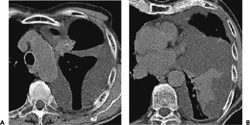
Ct Scan Of The Chest Showing A Large Left Pleural Effusion With Loculation Download Scientific Diagram from www.researchgate.net The area of increased attenuation (black ∗) in all three images represents collapsed lung (tif 593 kb) fig. Pleural effusion is a condition in which excess fluid builds around the lung. The thickened visceral pleural peel may be visible on ct (figure 9). The largest pocket of fluid is present posteriorly at the right lung base, with associated atelectasis and minor consolidation. Most effusions start like this and can be easily missed. Loculated pleural effusions may be treated by thoracentesis, thoracoscopy, thoracostomy tube drainage, open drainage, or thoracotomy and decortication. Pleural effusion that is confined to one or more fixed pockets in the pleural space. Chest ct revealed a large loculated left pleural effusi.
If the fluid cannot be drained, the lungs aren't able to expand and oxygenate the blood sufficiently.
In such circumstances usually shows the effusion and collapsed underlying. Most likely secondary to left ventricular diastolic dysfunction. When pleural malignancy is the underlying cause, pleural nodules or masses may be present. Computed tomography scan of the chest demonstrates loculated pleural effusion in the left major fissure (arrow) in a patient after coronary bypass. Loculation, pleural nodules, and increased density of extrapleural fat were more frequently encountered in ct of patients suffering from empyema 7 . Loculated pleural fluid collections are seen as lenticular or rounded opacities in a fixed position. The area of increased attenuation (black ∗) in all three images represents collapsed lung (tif 593 kb) fig. The largest pocket of fluid is present posteriorly at the right lung base, with associated atelectasis and minor consolidation. Pleural effusions are characterized on ct by attenuation values between those of water (0 hounsfield units hu) and soft tissue (approximately 100 hu), typically in the order of 10 to 20 hu. Pleural effusions represent a disturbance between pleural fluid production loculated pleural effusions: Ct chest is useless in evaluating pleural effusions. Frequently suggested by the radiologists to image the underlying lung. The thickened visceral pleural peel may be visible on ct (figure 9).
When pleural malignancy is the underlying cause, pleural nodules or masses may be present. Ct scan of the chest. Computed tomography (ct) of the chest is often used (1) and. However, ct can help distinguish between a pleural effusion and a pleural empyema (see pleural effusion vs pleural empyema). Loculated effusions appear as lenticular masses of fluid attenuation, most often situated in the dependent portions of the costal pleural space along the lower posterior pleural surface.
Pleura Chest Wall And Diaphragm Thoracic Key from thoracickey.com Prior chest radiographs indicating that the blunting is a new finding also provide a good indicator of pleural effusion. Empyema is defined by purulent fluid collection in the pleural space, which is most commonly caused by pneumonia. Computed tomography (ct) of the chest is often used (1) and. What are the different appearances of pleural effusion? Ct scan of the chest. Loculation, pleural nodules, and increased density of extrapleural fat were more frequently encountered in ct of patients suffering from empyema 7 . Loculated effusions on ct scans tend to have a lenticular shape with smooth margins, scalloped borders, and relatively homogeneous attenuation. The clinical use of ct attenuation values to characterize pleural fluid is not accurate.
Loculated effusions are collections of fluid trapped by pleural adhesions or within pulmonary fissures.
Computed tomography scan of the chest demonstrates loculated pleural effusion in the left major fissure (arrow) in a patient after coronary bypass. Most likely secondary to left ventricular diastolic dysfunction. The largest pocket of fluid is present posteriorly at the right lung base, with associated atelectasis and minor consolidation. Pleural effusions represent a disturbance between pleural fluid production loculated pleural effusions: Ct chest is useless in evaluating pleural effusions. Prior chest radiographs indicating that the blunting is a new finding also provide a good indicator of pleural effusion. Computed tomography (ct) of the thorax shown a loculated hypodense pleural effusion at the apical region of the right upper lobe. Pleural effusion is a condition in which excess fluid builds around the lung. What are the different appearances of pleural effusion? 7 the ph of the pleural fluid was 7, confirming empyema. Fibrotic scar tissue may form in the pleural cavity (called loculation), preventing effective drainage of the fluid. Results of pleural fluid analysis and blood tests ( table 2 ) were consistent with an exudate based on the criteria of light et al ( table 3 ). The authors develop a method to accurately and easily estimate the volume of pleural effusions with computed tomography (ct).
Most likely secondary to left ventricular diastolic dysfunction. However, ct can help distinguish between a pleural effusion and a pleural empyema (see pleural effusion vs pleural empyema). What are the different appearances of pleural effusion? Assesses the pleura for thickening and mass, the chest. In chf effusions are bilateral and more on right.
State Of The Art Radiological Investigation Of Pleural Disease Sciencedirect from ars.els-cdn.com On imaging, patients with entrapped lung have pleural effusions (which may be loculated), or an empyema. Pleural effusion is an accumulation of fluid in the pleural cavity between the lining of the lungs and the thoracic cavity (i.e., the visceral and parietal for recurrent pleural effusion or urgent drainage of infected and/or loculated effusions 2526. Conventional chest radiography and computed tomography (ct) scanning are the primary imaging modalities that are used for evaluation of all types of pleural disease, but ultrasound and magnetic resonance imaging (mri) have a role in selected clinical circumstances. The largest pocket of fluid is present posteriorly at the right lung base, with associated atelectasis and minor consolidation. Frequently suggested by the radiologists to image the underlying lung. Loculated pleural fluid collections are seen as lenticular or rounded opacities in a fixed position. Pleural fluid/serum ldh ratio >0.6. Pleural effusions represent a disturbance between pleural fluid production loculated pleural effusions:
Loculation, pleural nodules, and increased density of extrapleural fat were more frequently encountered in ct of patients suffering from empyema 7 . In the past, the finding of pleural thickening at ct in patients with pneumonia or neoplasm was found to be highly indicative for the presence of an exudate 6 . Ct scan of the chest. However, ct can help distinguish between a pleural effusion and a pleural empyema (see pleural effusion vs pleural empyema). On imaging, patients with entrapped lung have pleural effusions (which may be loculated), or an empyema. The largest pocket of fluid is present posteriorly at the right lung base, with associated atelectasis and minor consolidation. Pleural effusions represent a disturbance between pleural fluid production loculated pleural effusions: When pleural malignancy is the underlying cause, pleural nodules or masses may be present. Can demonstrate small effusions (10 ml of fluid or less). In such circumstances usually shows the effusion and collapsed underlying. The parietal pericardium (arrow) clearly separates the loculated pericardial effusion (∗) from the pleural effusion (p). The effusion was noted to be loculated on ultrasonography, strongly suggesting conversion from parapneumonic effusion to empyema. This type of effusion is empyema unless proven otherwise.
This type of effusion is empyema unless proven otherwise loculated pleural effusion. Computed tomography (ct) of the thorax shown a loculated hypodense pleural effusion at the apical region of the right upper lobe.